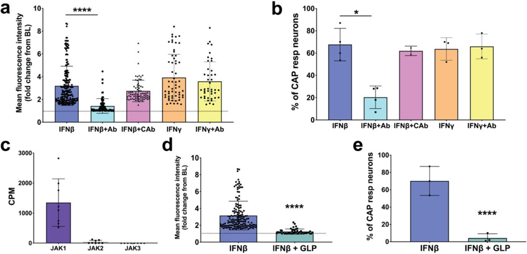
FIG 5: Type I Interferons activate airway nodose ganglia afferents via IFN Type I receptors signaling through JAK1 kinase.
a. Response of airway sensory nerves to IFNβ (1000 U/ml applied to the lungs via the trachea) in the absence and presence of IFN type 1 receptor blocking antibody (anti-IFNAR1, 1ug/ml applied 15 minutes before IFN treatment) or isotype control antibody; the response to IFN𝝲 (dose) in the absence and presence of the IFN1 blocking antibody is also shown. b. Graph showing the % of capsaicin (CAP) responsive neurons from experiments performed in panel a. c. Bar graph showing the presence of JAK1, JAK2 and JAK3 transcripts, measured as CPM, from nodose ganglia found using RNAseq. d. Graph comparing the mean fluorescence intensities after neuronal activation by IFNβ (1000 units/ml) in the absence and presence of the JAK1 inhibitor filgotinib/GLPG0634 (1μM) (dose perfused for 15 min before IFNβ) pretreatment to the airway afferents and then the application of IFN β. e. Graph showing % of CAP responsive neurons from experiments performed in panel d. (n=~400 neurons from n=4 mice for each treatment). All graphs show mean and SD. The asterisk denotes significant difference (* P< 0.05 and **** P< 0.0001) determined with unpaired One-way ANOVA test using GraphPad prism software (Bonferroni correction) was used throughout to compare unpaired groups.

