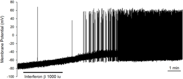
FIG 7: Direct application of IFNβ on the nodose ganglia and dissociated nodose ganglia neurons causes activation and action potential discharge.
IFNβ (1000U/ml) induced membrane depolarization (from −63.9±5 to −52.9±3.2 mV, n=7, P=0.009 by paired t-test) and action potential discharge in 4 out of 7 neurons with the mean peak frequency at 3.9±3.2 Hz. The tracing shown here is the membrane potential recording obtained from a capsaicin-sensitive nodose neuron using the perforated whole-cell patch clamp technique. The duration of IFNβ application by bath perfusion is indicated by the horizontal bar. The peak frequency of AP firing in this neuron is 9Hz.

