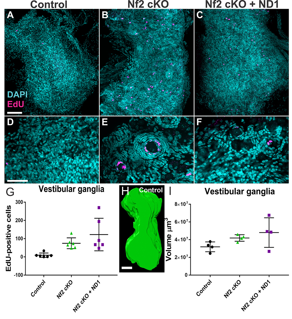
Figure 6.
Glial cell proliferation in control mice, mice with Nf2 cKO, and mice with Nf2 cKO+ND1. EdU uptake in adult mice aged 10–15 months was analyzed. Mice were injected with EdU (50mg/kg IP four times in a 24 hour period; 0, 4, 8 and 24hrs) and vestibular and geniculate ganglia were dissected within 72 hrs. EdU was detected in whole ganglion using the Click-It reaction and counterstained with DAPI. Ganglia were imaged with confocal microscopy and EdU quantified using the maximum projection of the z-stack. N=6 for each group. While only occasional spontaneous proliferations were found in control animals (A), both vestibular and geniculate intraganglionic glia cells showed a significant increase in proliferation that was not diminished by expression of Neurod1 (B,C). Details show that the proliferation in Nf2 cKO mice primarily occurs as an aggregation of flattened nuclei around a central neuron with the distribution of EdU positive cells nearest the neuron (E,F). Note that most Nf2 mutants had an abundance of these nodules which were nearly absent in control ganglia (D) and were smaller and fewer in Nf2 cKO+ND1 mice (compare B,C). Quantification of all EdU positive profiles in the entire vestibular and geniculate ganglia showed a clear difference between control and mutant mice (symbols reflect individual mice, vertical bar indicates standard deviation). While both Nf2 cKO and Nf2 cKO+ND1 mice had elevated levels of EdU compared to control, the latter group had the widest range of EdU positive cells that is in part related to overall size differences.

