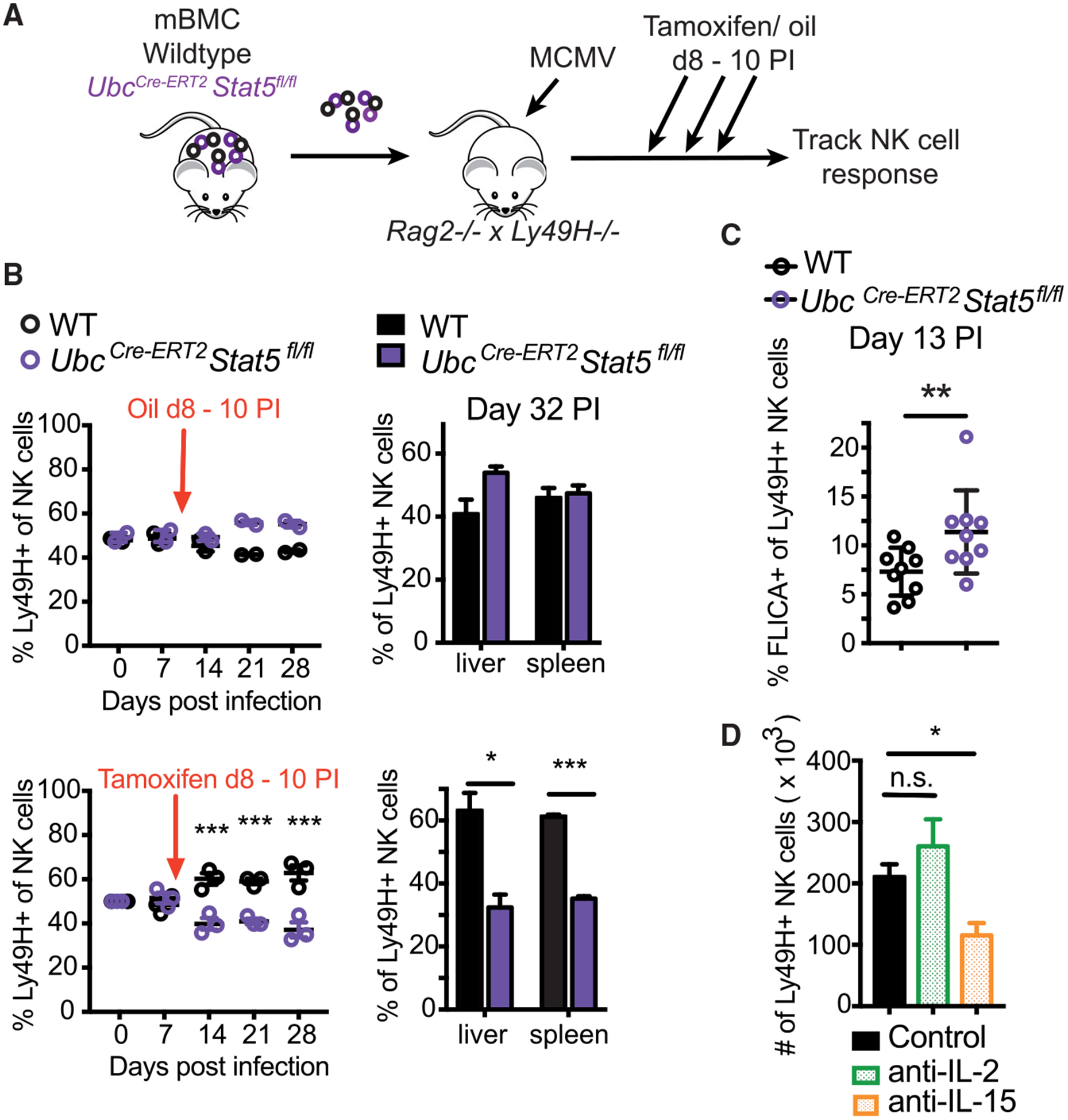
Figure 4. IL-15 and STAT5 Are Required for the Survival of Memory NK Cells.

(A–C) Splenic NK cells from WT:UbcCre-ERT2 × Stat5fl/fl mBMC mice were transferred into Rag2−/− × Ly49h−/− mice. Tamoxifen or oil (as a control) was administered on days 8 to 10 following MCMV infection. (B) Graphs show relative percentages of Ly49H+ WT and KO NK cells in the peripheral blood at indicated time points and in spleen and liver at day 32 PI. Data are representative of 2 independent experiments (n = 2–4). (C) Graph shows percentage of NK cells from spleen staining positive for FLICA on day 13 PI. Data are pooled from 2 independent experiments (n = 4–5).
(D) WT Ly49H+ NK cells were transferred into Ly49h−/− mice infected with MCMV, and recipient mice were treated with PBS, anti-IL-2, or anti-IL-15 from days 8 to 18 PI. Graph shows the number of NK cells in the spleen on day 30 PI. Data are representative of 2 independent experiments (n = 4–5).
All error bars indicate SEM.
