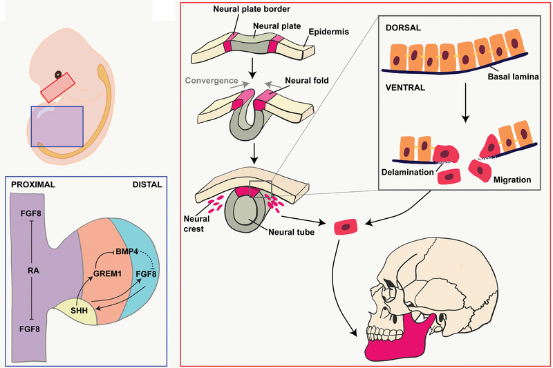
Figure 1. Embryonic View of Bone Development.
(A) Developing mandible (red box) and lower limb (blue box). (B) Endothelial-to-mesenchymal transition of cranial neural crest cells. (C) Signaling throughout the limb bud trunk (purple), zone of polarizing activity (yellow), progress zone (orange), and apical endodermal ridge (blue).

