
Figure 1. Morphologic features of RHE with YAP1 gene rearrangements.
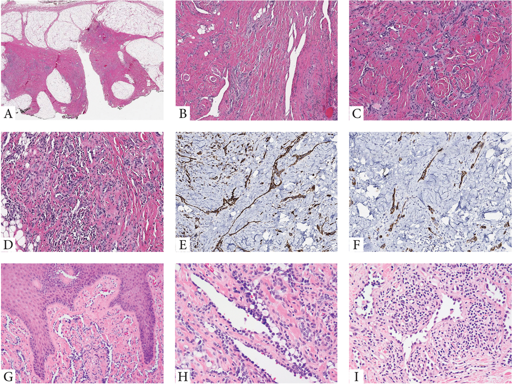
(A-F). Index case of a superficial soft tissue lesion from the knee in a 10 year-old male (case 1). Low power view showing a vague nodular and infiltrating growth pattern within subcutis which appears heavily fibrotic (A). Medium power shows densely sclerotic stromal background with either elongated, staghorn vessels (B), or shorter, retiform vascular network, lined by small endothelial cells with scant cytoplasm (C), with patchy prominent lymphocytic infiltrate, obscuring the vascular proliferation (D). Tumor cells typically co-express CD31 (E) and podoplanin (D2-40) (F). G-H. Cutaneous example showing elongated vascular channels infiltrating through the superficial dermis, lined by hobnailed endothelial cells (case 4, 10/M, buttock). I. Another RHE skin lesion in an adult (50/F, knee) showing angulated vessels with dilated lumina and protruding endothelial cells, some appearing detached and reminiscent of micropapillae. The vascular channels are embedded in a stroma with abundant lymphocytic infiltrate (case 5).