
Figure 4. Histologic features of the molecular negative subset of RHE and CHE.
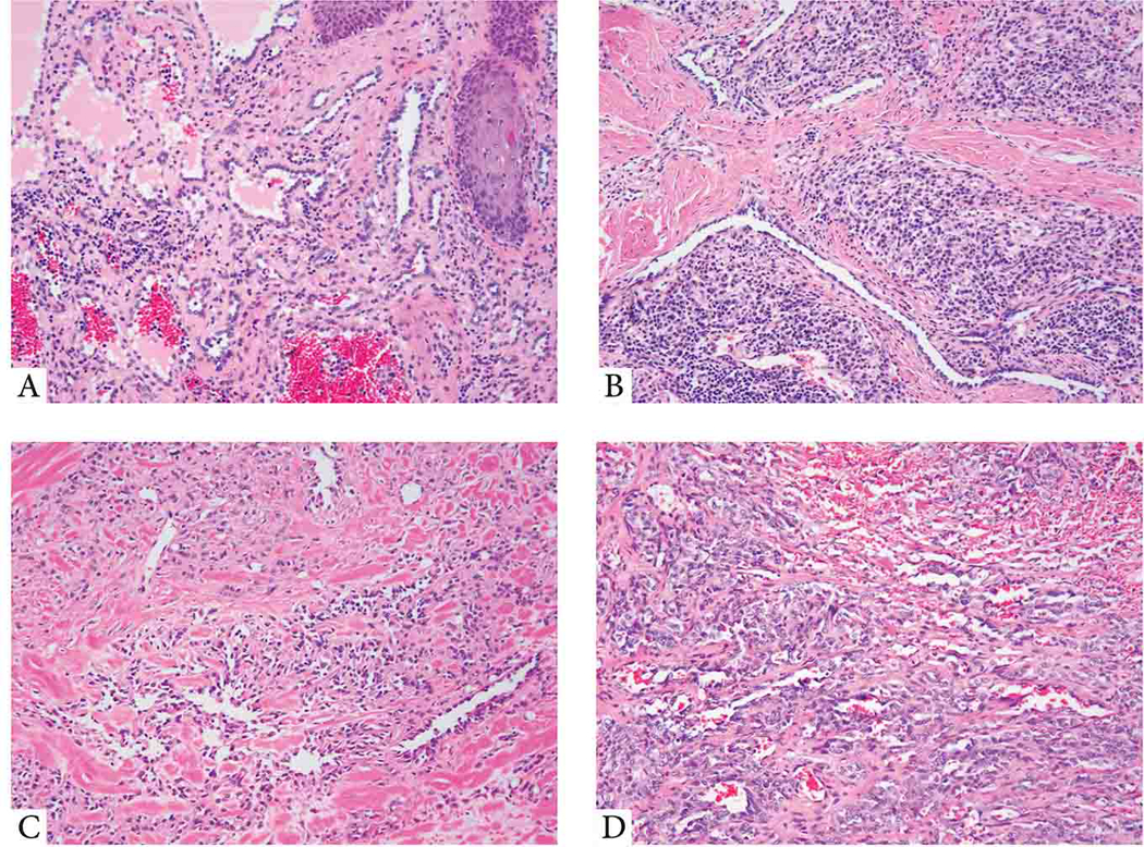
(A-B) YAP1-negative RHE. (A; 14/M, shoulder) Low power view showing dilated blood vessels within dermis lined by hobnail endothelial cells. (B; 55/M, thigh) Elongated, tubular vascular channels lined by cuboidal hyperchromatic endothelial cells, embedded in a stroma with abundant lymphocytic infiltrate. (C,D) CHE lacking YAP1 or MAML2 gene rearrangements. (C; 24/F, scalp) Predominant retiform areas with a focal epithelioid component arranged in single files. (D; 36/F, scalp) A variegated appearance showing solid, single cells as well as hemangioma-like areas.