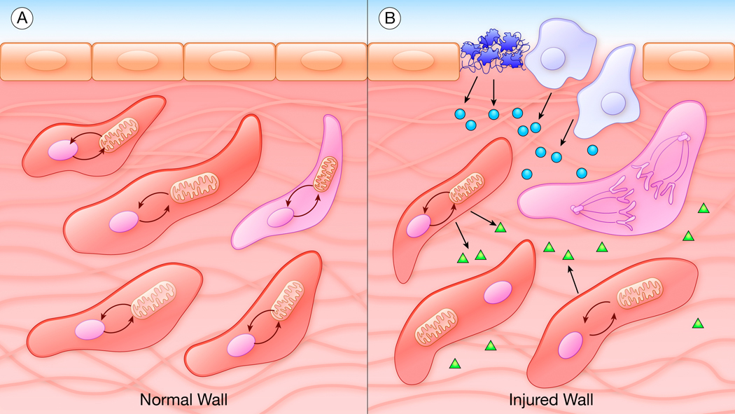
Figure.
Metabolic-transcriptional coupling in smooth muscle cells (SMCs). A, In the normal vessel wall, SMCs appear homogeneous but actually exist in many different real or potential phenotypes based on local variations in metabolic reactions and their links to transcriptional coupling. B, In the injured vessel wall, preexisting states of metabolic-transcriptional coupling mean that only a small number of “primed” SMCs are able to cross a critical threshold and respond to the change in environment. Platelets (dark blue), leukocytes (light blue), endothelial cells (tan), secreted factors (circles, triangles), SMCs (red), primed and dividing SMC (pink).

