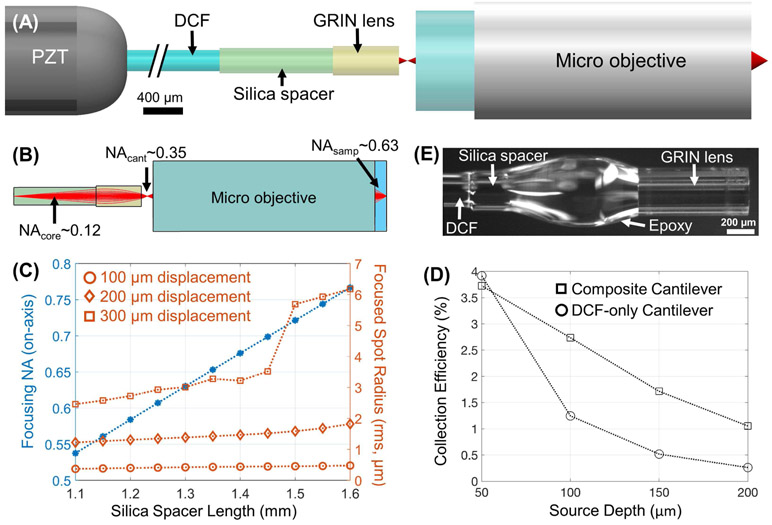
Fig. 1.
Design of the composite cantilever. (A) Schematic of the composite cantilever paired with a micro objective. (B) Illustration of the evolving beam NA from the DCF tip (and within the spacer), to the new beam focus near the exit surface of the GRIN rod lens, and the final beam focus in the sample (water immersion). (C) The dependence of the on-axis focusing NA and off-axis focused spot radii (root-mean-square, at three progressive tip displacements) on the silica spacer length extracted from ray-tracing simulations. For simplicity, we did not consider the angular deflection of the beam exiting the cantilever in our ZEMAX simulations since the deflection angle was very small (e.g., no larger than 1.7 degrees for both the long and short cantilevers) in comparison to the ~0.35 beam NA exiting the composite cantilever. (D) Collection efficiency comparison between two endomicroscope designs: 1) a traditional DCF only cantilever and 2) the proposed composite cantilever. Each data point shown here is the average value over 5 different scattering anisotropy factors. (E) Photograph of a finished composite cantilever tip.

