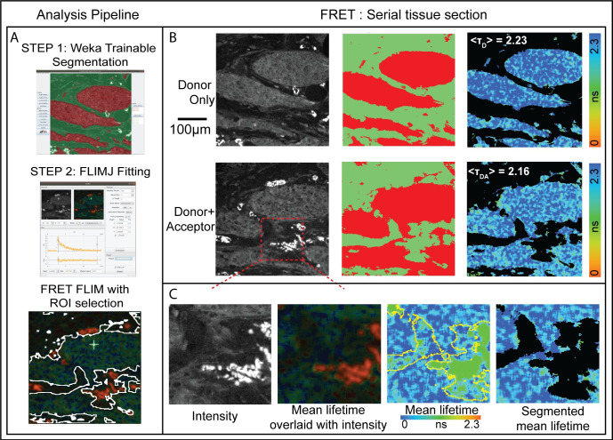
Fig 2.
A) Breast cancer tissues from 107 patients METABRIC study were stained with antibodies: anti-HER3-IgG-Alexa546 (donor) and anti-HER2-IgG-Cy5 (acceptor) [37]. Serial sections were stained with donor+acceptor (DA, FRET pair) and with donor alone (D, control). Average lifetime values (TD and TDA) can be determined for the tumor from the two serial sections using FLIMJ after segmentation. The FRET efficiency can be calculated according to FRETeff = 1 –TDA/TD. B) Weka Trainable Segmentation plugin was used to segment the tissue areas. The FLIMJ user interface showing a typical transient and fit from the tissue. We used the LM fitting with a mono-exponential model. C) Zoom into a smaller region. Composite image from FLIMJ showing lifetime information. Pure lifetime map with Weka segmentation shown in yellow. Segmentation result of the lifetime within the tumor with artifactual tissue removed. From TD = 2.23 ns and TDA = 2.16 ns, we estimate a FRET efficiency for this example tumor area as 3.1% as a measure of HER2-HER3 dimerization on the tumor in this patient.

