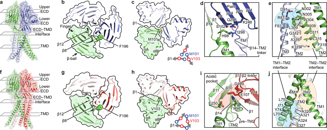
Figure 2: Intersubunit interfaces.
Each column represents the same view of pH8–PAC (upper) and pH4–PAC (lower). a and f, The overall structure of PAC shown in cartoon and surface representation. The extracellular domain (ECD) is divided into the upper ECD and lower ECD for discussion. b and g, The upper ECD viewed from the extracellular side. F196, which mediates the intersubunit interaction in the upper ECD, is shown as spheres. c and h, The lower ECD viewed from the extracellular side. At pH 8, M101 at the beginning of the β1 strand is in the center of lower ECD (lower right of panel c). At pH 4, the lower ECD undergoes a clockwise inward rotation so that V103 in the middle of the β1 strand moves to the center of the lower ECD (lower right of panel h). d and i, The ECD–TMD interface viewed parallel to the membrane. e and j, The interaction interfaces at the TMD.

