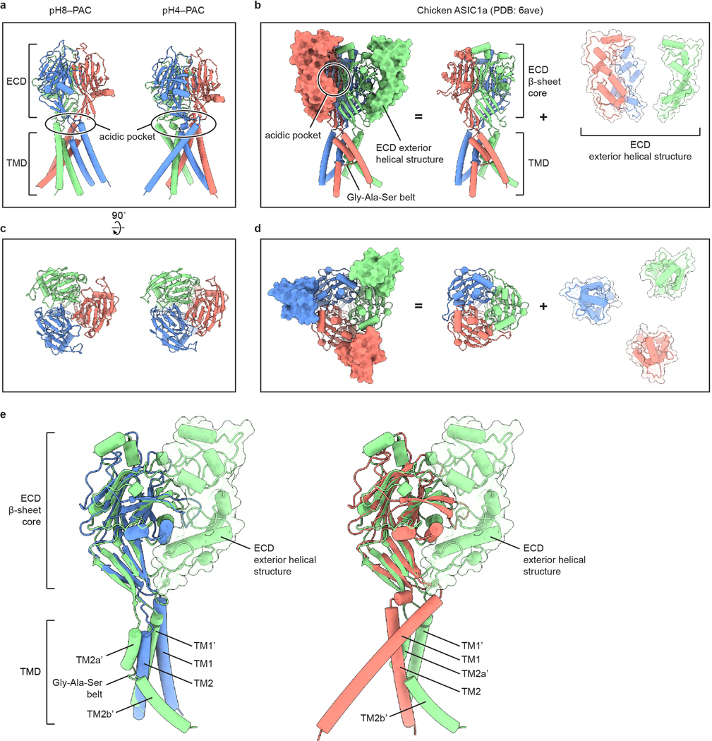
Extended Data Figure 5: Comparison of the structures of PAC and ASIC.

a-d, Structural comparison of human PAC (a, c) with chicken ASIC1a (b, d) viewed parallel to the membrane (a, b) and from the extracellular side (c, d). The acidic pocket of human PAC and chicken ASIC1a are in different locations. e, Overlay of the pH8–PAC (blue) and pH4–PAC (red) single subunit with the chicken ASIC1a (green) subunit. The ECD of ASICa is composed of a β-sheet core and the exterior helical structure. While the β-sheet core shares high similarity to human PAC structure, the chicken ASIC1a TMD is organized differently from that of the human PAC.
