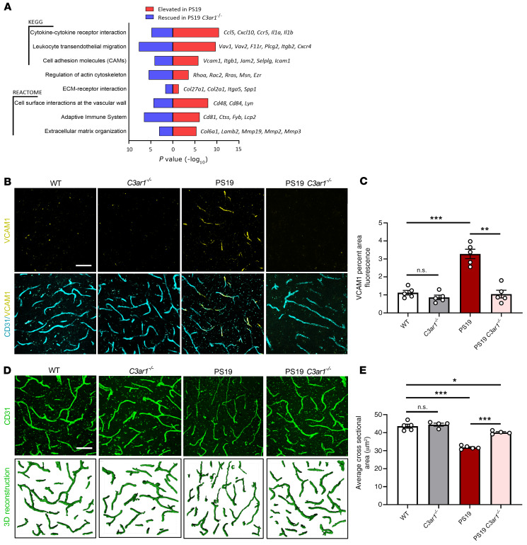
Figure 8. Vascular abnormalities in PS19 tau–transgenic mice and C3aR dependency.
(A) RNA-Seq analysis revealed significantly overrepresented pathways in the differentially expressed genes (DEGs) that were increased in 9-month-old PS19 compared with WT animals (red), and that were decreased in PS19 C3ar1–/– compared with PS19 animals (blue). Terms were selected from results based on their involvement in vascular biology and immune cell infiltration and plotted by P value; representative rescued genes contributing to the terms are listed (right). (B) Cortical staining of 9-month-old WT, C3ar1–/–, PS19, and PS19 C3ar1–/– hippocampal vasculature with CD31 and VCAM1 demonstrated a significant increase in VCAM1 expression in PS19 mice and a rescue of this phenotype in PS19 mice harboring the C3ar1 deletion. (C) Quantification of VCAM1 immuno-intensity. (D) CD31 staining and IMARIS-aided 3D reconstruction of 9-month-old WT, C3ar1–/–, PS19, and PS19 C3ar1–/– hippocampal vasculature. (E) Quantification of the average vessel cross-sectional area. All data represent the mean ± SEM of n = 5/group. Analysis for all results was performed using 1-way ANOVA with Tukey’s post hoc test (*P < 0.05, **P < 0.01, ***P < 0.001). Scale bar: 50 μm.

