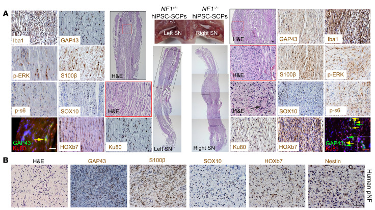
Figure 3. NF1–/– hiPSC-SCPs give rise to pNFs.
(A) The non–tumor-bearing left sciatic nerve (SN) injected with NF1+/– hiPSC-SCPs and the neurofibroma-bearing right sciatic nerve injected with NF1–/– hiPSC-SCPs were fully characterized by H&E staining as well as immunostaining with the human-specific Ku80, GAP43, S100β, SOX10, HOXb7, p-ERK, p-s6, and Iba1 antibodies. Yellow arrows show the colocalization of GAP43+ and Ku80+ cells. Green arrows show GAP43+Ku80– cells. Black arrow shows the Meissner-like corpuscle in the neurofibroma. n = 5. (B) Characterization of human pNF tissue by H&E staining and immunostaining for GAP43, S100β, SOX10, HOXb7, and nestin. SN, sciatic nerve. Scale bars: 50 μm and 25 μm (inset in A).

