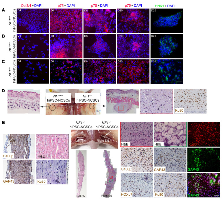
Figure 4. The nerve microenvironment promotes NCSC differentiation into SLCs and the formation of neurofibromas.
(A–C) After differentiation, hiPSC-NCSCs were immunonegative for Oct3/4 on day 4, immunopositive for p75 on days 4, 8, and 20, and immunopositive for HNK1 on day 20. (D) hiPSC-NCSCs were subdermally injected into athymic mice. Formation of cartilage derived from injected Ku80+ cells was observed under the skin following NF1–/– hiPSC-NCSC implantation, but not in the left sciatic nerve after NF1+/+ hiPSC-NCSC implantation. n = 3. (E) hiPSC-NCSCs were injected into the sciatic nerves of athymic mice. Formation of cartilage and tumors with neurofibroma histological and molecular characteristics was observed in the right sciatic nerves following implantation of NF1–/– hiPSC-NCSCs. Colocalization of Ku80 and GAP43 was observed. The left sciatic nerve injected with NF1+/+ hiPSC-NCSCs was immunopositive for Ku80 but still well-organized, without histological features of neurofibroma. n = 3. White arrow points to tumor in the right sciatic nerve. Scale bars: 50 μm and 10 μm (inset in E).

