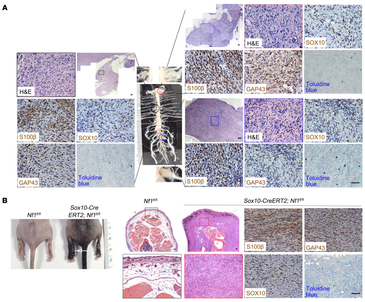
Figure 7. SOX10-expressing cells contain proliferating tumorigenic cells for pNF.
(A) Sox10-CreERT2 Nf1fl/fl mice treated with tamoxifen demonstrated neurofibroma formation, characterized by abnormally enlarged DRGs, as well as hypercellular and disorganized DRGs. The pNF was positive for S100β, GAP43, and SOX10 expression, with infiltration of mast cells. (B) A representative Sox10-CreERT2 Nf1fl/fl mouse treated with tamoxifen developed classic giant, diffuse pNFs (white arrow) with hyperpigmentation and thickening of the skin, which was positive for S100β, GAP43, and SOX10 expression, with mast cell infiltration. n = 43. Scale bars: 50 μm.

