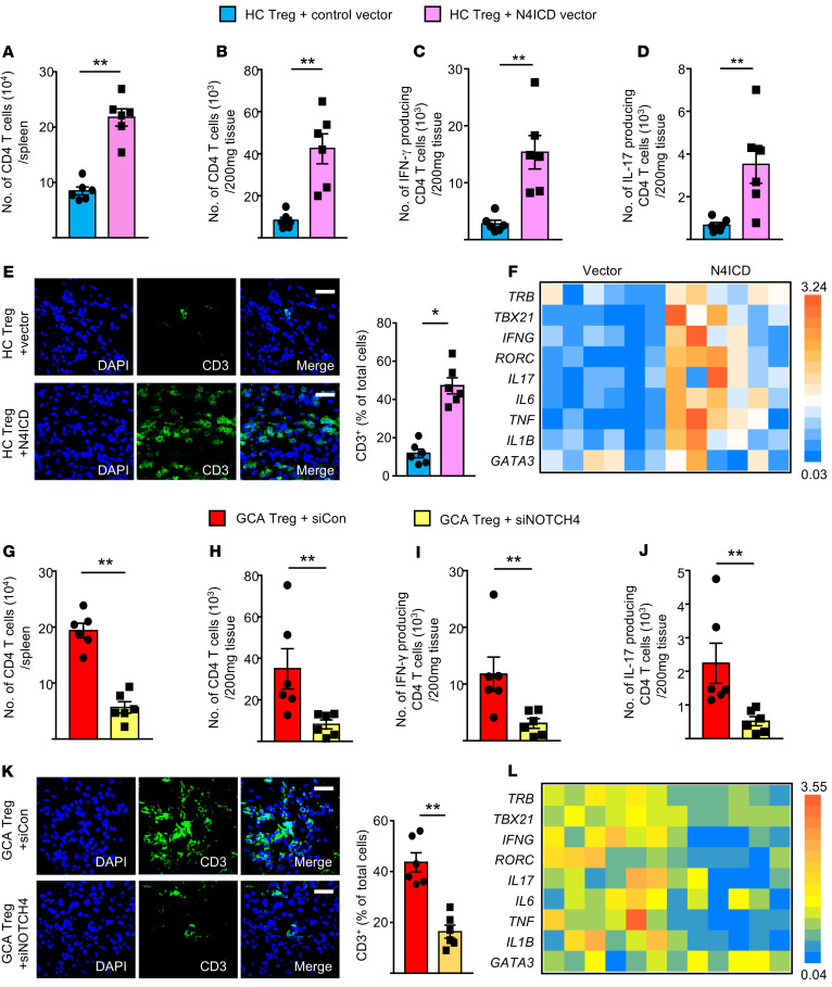
Figure 3. NOTCH4 signaling promotes vascular inflammation.
To induce vasculitis, NSG mice were engrafted with human arteries and reconstituted with a mixture of PBMCs and CD8+ Tregs as in Figure 1. Prior to the adoptive transfer, NOTCH4 signaling in healthy, induced CD8+ Tregs was enhanced by transfecting them with an N4ICD-containing vector (A–F). Alternatively, NOTCH4 signaling in patient-derived CD8+ Tregs was suppressed by siRNA transfection (G–L). Human arteries were explanted and examined by IHC staining, tissue transcriptome analysis, and flow cytometry of extracted T cells. (A and B) Numbers of human CD4+ T cells in the spleen and in the engrafted artery analyzed by FACS. (C and D) Tissue-infiltrating T cells were isolated from the inflamed arteries, and IFN-γ and IL-17 production were assessed by intracellular staining and flow cytometry. (E) Tissue-infiltrating T cells in the arteries analyzed by immunostaining for CD3 (green). Nuclei marked with DAPI. Representative images from 6 grafts. Scale bars: 20 μm. (F) Tissue transcriptome in explanted arteries analyzed by RT-PCR. (G and H) Numbers of human CD4+ T cells in the spleen and in the human arteries analyzed by FACS. (I and J) Flow cytometric staining of intracellular IFN-γ and IL-17 in T cells extracted from explanted human arteries. (K) Tissue-infiltrating T cells in the arteries analyzed by immunostaining for CD3 (green). Nuclei marked with DAPI. Representative images from 6 grafts. Scale bars: 20 μm. (L) Tissue transcriptome analysis in explanted arteries by RT-PCR. All data are mean ± SEM from 6 samples. *P < 0.05; **P < 0.01 by paired Mann-Whitney-Wilcoxon rank test.

