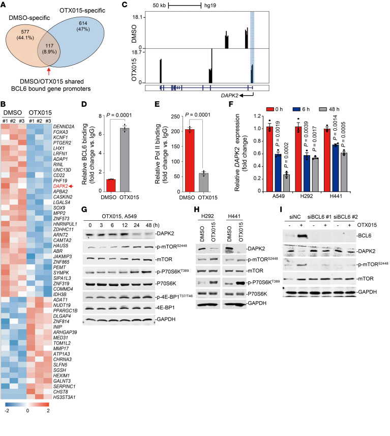
Figure 5. Increased BCL6 activates the mTOR pathway through suppression of DAPK2.
(A) Venn diagram shows the overlap of BCL6-bound gene promoters between the DMSO and OTX015 treatment groups. These data were generated by the ChIP-Seq experiments described in Figure 3A. (B) Heatmap of differentially expressed genes (fold change >2, adjusted P < 0.05) in the OTX015-specific region described in A. RNA-Seq was performed using the same cell samples as the ChIP-Seq in Figure 3A. Blue represents gene downregulation, and red represents gene upregulation, with 0 as the median. (C) Genome Browser tracks showing BCL6 ChIP–Seq signals in the DAPK2 gene locus in A549 cells treated with DMSO or OTX015 (300 nM) for 6 hours. Blue shading marks the peaks located in the promoter region. (D) BCL6 binding level at the promoter region of DAPK2 examined by ChIP-qPCR assays. (E) RNA Pol II–binding level at the promoter region of DAPK2 examined by ChIP-qPCR assays. Data represent the mean ± SEM of biological triplicates. The P value was determined by comparing the OTX015 treatment group with the control group, using an unpaired, 2-tailed Student’s t test. (F) Relative DAPK2 expression at different time points. Cells were treated with 300 nM OTX015, and DAPK2 mRNA levels were detected by qPCR. Data represent the mean ± SEM of 3 independent experiments. P values were determined by comparing OTX015-treated groups with the untreated group, using an unpaired, 2-tailed Student’s t test. (G) Effects of OTX015 on the DAPK2/mTOR signaling pathway on A549 cells. A549 cells were treated with OTX015 (300 nM) for the indicated durations. Treated cells were collected and probed with antibodies against DAPK2, p-mTOR (Ser2448), mTOR, p-P70S6K (Thr389), P70S6K, p–4E-BP1 (Thr37/46), 4E-BP1, and GAPDH, respectively. (H) Effects of OTX015 on the DAPK2/mTOR signaling pathway in H292 and H441 cells. (I) BCL6 knockdown impaired OTX015-mediated DAPK2 suppression and mTOR activation. BCL6 silencing was conducted by RNA interference in A549 cells. Cells were treated with 300 nM OTX015 for 48 hours. Cells lysates were collected and probed with antibodies against BCL6, DAPK2, p-mTOR (Ser3448), mTOR, and GADPH. Immunoblots in G, H, and I were contemporaneous and run in parallel from the same biological replicate, respectively. The immunoblots are representative of at least 3 independent experiments.

