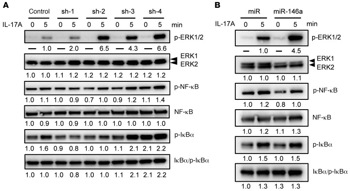
Figure 4. Reduced galectin-7 expression and miR-146a overexpression promote ERK1 and ERK2 signaling pathways triggered by IL-17A.
(A) Galectin-7–knockdown HaCaT cells and control cells were treated with IL-17A for 5 minutes, and cell lysates were analyzed by immunoblotting. Total ERK1, ERK2, NF-κB, and IκBα and their phosphorylated forms were detected with the corresponding antibodies. (B) HaCaT cells stably transfected with pmiR (control vector) or pmiR-146a were treated with IL-17A for 5 minutes. Immunoblotting was performed as described in A. Protein quantification data on phospho-ERK1 (p-ERK1) and phospho-ERK2 (p-ERK2) were normalized to the control group in A and the miR group in B. Data on total protein levels, levels of phosphorylated NF-κB and IκBα, and total protein levels of ERK1 and ERK2 were normalized to the control (0 minutes).

