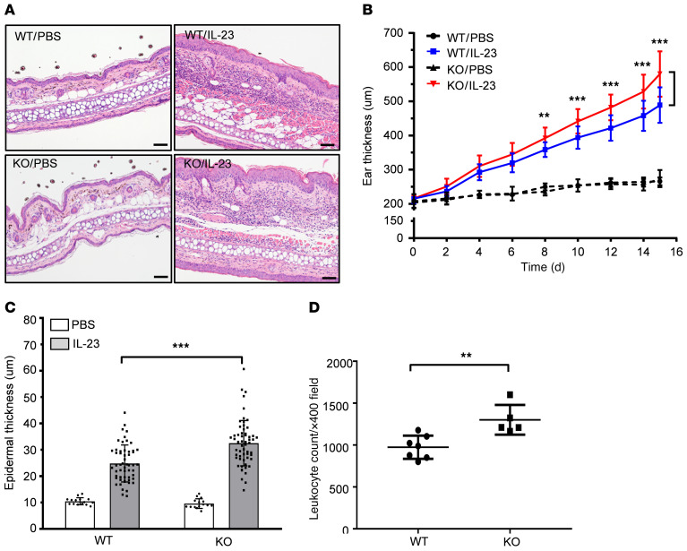
Figure 5. Galectin-7–deficient mice exhibit hyperproliferative keratinocytes and increased leukocyte infiltration at the intradermally IL-23–injected sites, as compared with their littermate controls.
(A) H&E staining of ear sections from WT or galectin-7–deficient (knockout; KO) mice injected intradermally with PBS or IL-23 every other day for 15 days. Scale bars: 50 μm. (B) Ear thickness of WT and galectin-7–deficient (KO) mice was measured before each intradermal injection, and measurements were taken at the center of the ears (WT/PBS, n = 5; WT/IL-23, n = 18; KO/PBS, n = 5; KO/IL-23, n = 19). For statistical analysis, ear thickness of KO/IL-23 was compared with that in the corresponding WT/IL-23 group at each time point. (C) Epidermal thicknesses of WT and KO mice from H&E-stained sections as described in A obtained on day 15 from the same mice as described in B. For each tissue section, 3 measurements were taken. (D) Leukocytes were counted in ×400 magnified visual fields of tissue sections from IL-23–injected mice using the cell counting module in ImageJ software (WT, n = 7; KO, n = 5). All results are presented as mean ± SD. For statistical analysis, 2-way ANOVA with Tukey’s multiple-comparison test (B and C) or unpaired Student’s t test (D) was performed. **P < 0.01, ***P < 0.001.

