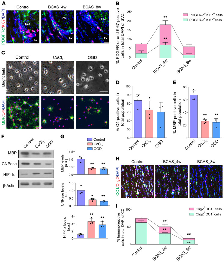
Figure 2. Pathological changes to OPCs and mature oligodendrocytes after hypoxic-ischemic injury.
(A and B) Changes in endogenous OPC numbers and their proliferation represented by Ki67+PDGFR-α+ cells as a percentage of total DAPI+ cells in the subventricular zone (SVZ) (n = 4 in each group). (C–E) Light microscopic appearance and fluorescence images displaying the differences in OPC differentiation and its quantification (n = 4 experiments). (F and G) Immunoblotting analyses of MBP and CNPase expression showed that hypoxia induced by CoCl2 (10 μM) or OGD (2 hours) impairs OPC maturation (n = 3 experiments). (H and I) Immunofluorescence images and quantification of the numbers of CC1+Olig2+ cells in the CC of BCAS mice compared with controls (n = 3 in each group). Data are presented as mean ± SD. *P < 0.05, **P < 0.01 vs. controls by 1-way ANOVA with Tukey’s post hoc test. Scale bars: 20 μm.

