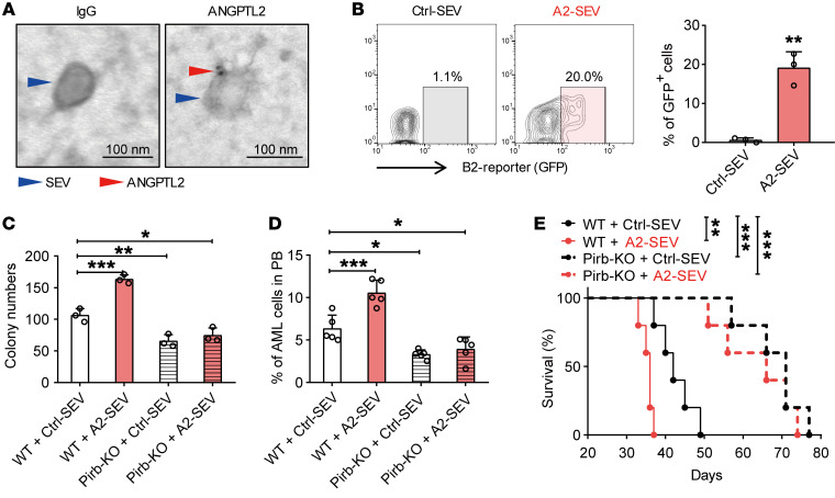
Figure 8. ANGPTL2-SEVs bind to LILRB2.
(A) Immunoelectron microscopy showing the location of ANGPTL2 protein in SEVs. Scale bars: 100 nm. (B) Flow cytometry (left) and histogram (right) analysis of ANGPTL2-SEVs binding to LILRB2-chimeric reporter cells. In this experiment, LILRB2-chimeric reporter cells will express GFP when ANGPTL2-SEVs bind to LILRB2 on the cell surface (n = 3; the data represent the means ± SD; **P < 0.01, Student’s t test). The stable chimeric receptor reporter cell system has been previously described (49). The extracellular domain of LILRB2 was chimerically fused with the intracellular domain of paired immunoglobulin-like receptor (PILR). Upon binding with ANGPTL2-SEVs, the receptor can further recruit the adaptor DAP-12 to activate the NFAT promoter, followed by the increase in GFP expression in reporter cells. (C) Colony-forming ability of Pirb+/+ and Pirb–/– AML cells cocultured with control- or ANGPTL2-SEVs (10 μg per 50,000 AML cells) (n = 3; the data represent the means ± SD; *P < 0.05, **P < 0.01, ***P < 0.001, 1-way ANOVA followed by Dunnett’s test). (D) The percentage of AML cells in the PB of recipients injected with Pirb+/+ and Pirb–/– AML cells cocultured with control- or ANGPTL2-SEVs (n = 5; the data represent the means ± SD; *P < 0.05, ***P < 0.001, 1-way ANOVA followed by Dunnett’s test). (E) Survival curves of recipients injected with Pirb+/+ and Pirb–/– AML cells cocultured with control- or ANGPTL2-SEVs (n = 5; **P < 0.01, ***P < 0.001, log-rank test). Experiments were conducted 2–4 times for validation.

