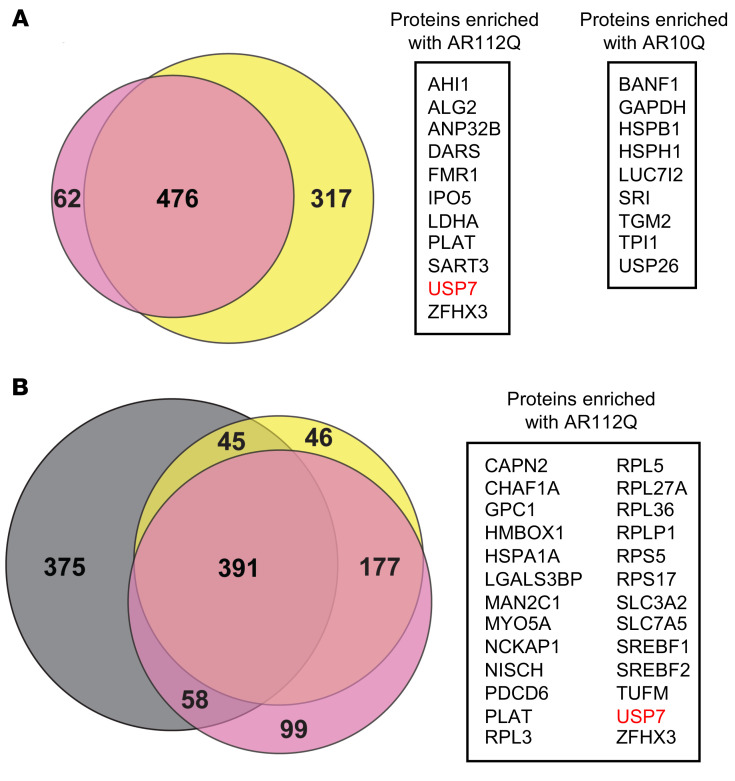
Figure 1. Unbiased quantitative interaction screen for WT and polyQ-expanded AR.
Three independent SILAC experiments were performed. Experiment 1: PC12 cells expressing AR 112Q or AR10Q were grown in heavy or light SILAC medium, respectively (shown in pink). Anti-AR (A) or 3B5H10 antibody (B) was used for IP. Experiment 2 (label swap): AR112Q- or AR10Q-expressing cells were grown in light or heavy SILAC medium, respectively (shown in yellow). Anti-AR (A) or 3B5H10 antibody (B) was used for IP. Experiment 3: cells expressing AR 112Q or AR10Q were grown in heavy or light SILAC medium, respectively (shown in gray). 3B5H10 antibody (B) was used for IP. Cells were induced with doxycycline to express AR and treated with DHT for 48 hours. (A) Left: Venn diagram comparison of common proteins pulled down with anti-AR antibody in 2 experiments. Right: common proteins that were enriched either with AR112Q or AR10Q by 1.5-fold or more. Fold enrichment is shown in Supplemental Tables 1 and 2. (B) Left: Venn diagram comparison of common proteins pulled down with 3B5H10 antibody in 3 independent experiments. Right: common proteins enriched with AR112Q by 1.5-fold or more in at least 2 experiments. Fold enrichment is shown in Supplemental Table 1.

