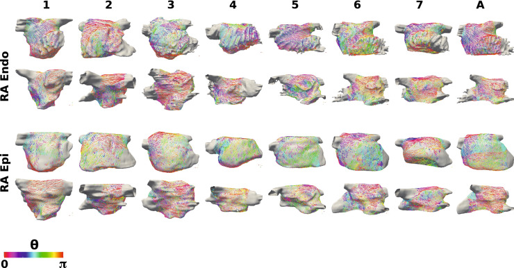
Figure 5.
Right atrial fibre fields. Right atrial body fibre fields are displayed as streamlines coloured by UAC angle for the endocardial (top two rows) and epicardial (bottom two rows) surfaces. These are shown in lateral-septal view (first and third row) and septal-lateral view (second and fourth row). These are orientated such that the IVC-SVC universal atrial coordinate axis is aligned with the x-axis and the lateral-septal coordinate is aligned with the y-axis. UAC angles of 0 or correspond to the horizontal direction (aligned with the IVC-SVC coordinate axis), and UAC angles of correspond to the vertical direction (aligned with the lateral-septal coordinate axis). Streamlines originating from the SVC, IVC, RAA and CS are omitted for visualisation purposes.

