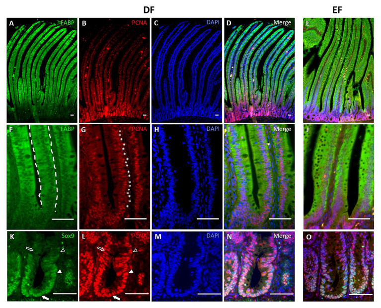
Figure 4.
Stem, progenitor, proliferating, and differentiated cells exhibit specific localization within villi and crypts at d10. IF of enterocyte marker FABP, PCNA, DAPI nuclear staining, and their merge at X100 magnification (A–D), and X400 magnification (F–I), and stem/progenitor cell marker Sox9, PCNA, DAPI, and their merge (K–N) in DF chicks at d10. Merged immunofluorescent images of the same markers in EF chicks at d10 are presented in the right, separate panel: FABP and PCNA at a magnification of X100 (E) and X400 (J); Sox9 and PCNA at X400 magnification (O). FABP expression was localized to all villus cells (F, dashed outlines). Proliferating cells were found along the villus (G, asterisks), colocalizing with FABP+ cells. Sox9 expression was restricted to crypts (K). Arrows indicate Sox9+/PCNA− cells, arrowheads indicate Sox9+/PCNA+ cells, arrow outlines indicate Sox9−/PCNA+ cells, and arrowhead outlines indicate Sox9−/PCNA− cells (K,L).

