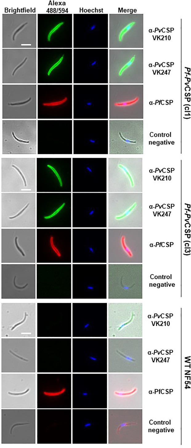
Figure 2.
Immunofluorescence analyses of PvCSP and PfCSP expression in fixed Pf-PvCSP salivary gland sporozoites. Fixed permeabilized sporozoites of Pf-PvCSP (clone 1 and clone 3) and WT PfNF54 parasites stained with mouse anti-PvCSPVK210, anti-PvCSP VK247 (green; alexa 488) and anti-PfCSP antibodies (red; alexa 594). Nuclei stained with the Hoechst-33342. Control negative, corresponds to the incubation without the primary antibody (2 experiments). Scale bar is 5 µm.

