FIG 2.
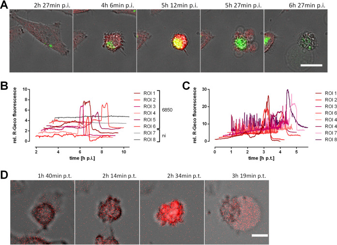
Infection with S. aureus or treatment with secreted S. aureus virulence factors leads to a massive cytosolic Ca2+ increase in epithelial cells. HeLa R-Geco cells were either infected with S. aureus 6850 green fluorescent protein (GFP) (A, B) or treated with 10% sterilized supernatant (SNT) of a S. aureus 6850 overnight culture (C, D) and visualized by live cell imaging. (A) Stills from time-lapse imaging are shown (green, S. aureus; red, R-Geco; gray, brightfield). Bar, 25 μm. (B) Quantification of relative R-Geco fluorescence of single cells (region of interest [ROI]) infected with S. aureus 6850 or not infected (ni) was performed over the course of infection. h p.t., hours posttransduction. (C) Relative R-Geco fluorescence of single cells was quantified upon SNT treatment. (D) Representative stills of one SNT-treated HeLa R-Geco cell (red, R-Geco; gray, brightfield). Bar, 10 μm.

