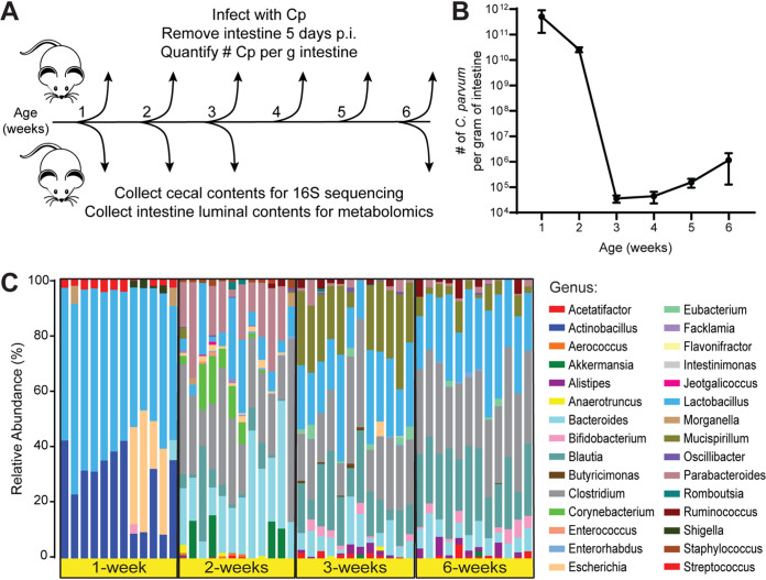
FIG 1.
Differences between C. parvum infectivity and cecal microbiota during murine postnatal development. (A) Diagram of the experimental design. Separate cohorts of mice were challenged with 5 × 10⁴ C. parvum oocysts at each week of life (n = 5 to 10 mice per week). Five days postinfection (p.i.), intestines were removed, and the number of C. parvum organisms per gram of intestine was quantified by qPCR. In a separate experiment, cecal contents and small intestinal luminal contents were collected from uninfected mice at 1, 2, 3, and 6 weeks of age (n = 12 mice per week) for 16S rRNA sequencing and metabolomics, respectively. (B) Line graph depicting the average number of C. parvum organisms per gram intestine of mice infected at the indicated weeks of age (mean ± SD, n = 10 mice each for weeks 1 and 2, n = 5 mice each for weeks 3 to 6). (C) Taxonomic differences in the cecal microbiota of mice at 1, 2, 3, or 6 weeks of age displayed as a stacked bar graph of the relative abundances of the bacterial genera detected by 16S rRNA sequencing.

