FIG 3.
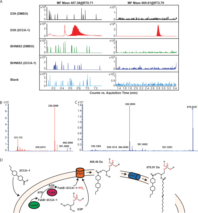
Lipidomic analysis of 2CCA-1 treated pneumococci. (A) Chromatograms showing molecular features (MF) 407.38 and 669.61 in wild-type Tigr4 in comparison with the fakB3 mutant BHN852, when treated with 2CCA-1 (25 μM) or DMSO (1% [vol/vol]) as a solvent control. The prominent peaks of MF 407.38 at retention time (RT) 0.71 min and MF 669.61 at RT 2.79 min were found only in 2CCA-1-treated wild-type Tigr4 but were absent in the DMSO-treated Tigr4 sample as well as in the spontaneous resistant fakB3 mutant BHN852. The color spikes in other samples are regarded as background noise (observe difference in scale [104] in the 2CCA-1-treated wild-type Tigr4 sample). MS-MS spectra of (B) MF 407.38, RT 0.71 min, and (C) MF 669.61, RT 2.79 min, show similar fragments (320.29 and 391.36), indicating similar molecular structures. (D) Proposed model for 2CCA-1-mediated toxicity. 2CCA-1 associates with the pneumococcal plasma membrane, where it is taken up by FakB3 and gets phosphorylated by FakA, to become a substrate for PlsY. PlsY acylates 2CCA-1 onto glycerol-3 phosphate (G3P) to form lysophosphatidic acid (with a calculated mass of 406.46 Da). The subsequent addition of an 18 carbon unsaturated fatty acid by PlsC forms a 2CCA-1-containing phosphatidic acid (with a calculated mass of 670.91 Da).

