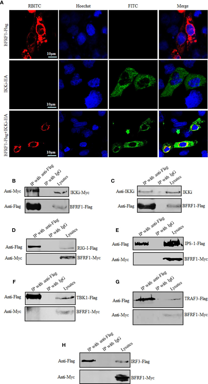
Figure 5.

BFRF1 co-localizes and interacts with IKKi. (A) COS-7 cells were transfected with expression plasmid pIKKi-HA or BFRF1-Flag, or co-transfected with plasmids combination pIKKi-HA and BFRF1-Flag. At 24 h post-transfection, IFA analysis was performed with primary Abs anti-HA and anti-Flag mAb, and their corresponding fluorescent secondary Abs FITC-conjugated goat anti-mouse IgG (green) and RBITC-conjugated goat anti-rabbit IgG (red), respectively. Cells were counterstained with Hoechst to visualize the nuclear DNA (blue) for 5 to 10 min. Images were obtained by confocal microscopy using a 63× lens objective. All scale bars indicate 10 μm. (B, C) HEK293T cells were transfected with plasmid BFRF1-Flag (C) or co-transfected with plasmids combination BFRF1-Flag and pIKKi-Myc (B). At 24 h post-transfection, cells were lysed and immunoprecipitated with anti-Flag mAb or mouse nonspecific IgG, then WB analysis was performed using the indicated Abs. (D–H) HEK293T cells were co-transfected with plasmids combination of pRIG-I-Flag/pBFRF1-Myc (D), pIPS-1-Flag/pBFRF1-Myc (E), pTBK1-Flag/pBFRF1-Myc (F), pTRAF3-Flag/pBFRF1-Myc (G), or pIRF3-Flag/pBFRF1-Myc (H). At 24 h post-transfection, cells were lysed and immunoprecipitated with anti-Flag mAb or mouse nonspecific IgG, then WB analysis was performed using the indicated Abs.
