
Fig. 3.
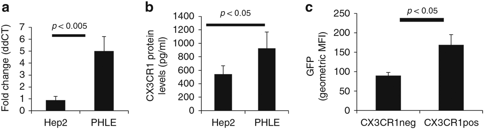
Role of CX3CR1 in RSV infection of pediatric epithelial cells. a RNA transcript levels of PHLE cell cultures 14 days post-ALI (N = 4) measured by quantitative PCR for the CX3CR1 transcript. HEp-2 cell line (N = 3) was used for comparison. b Protein levels of PHLE cell cultures 14 days post-ALI (N = 4) measured by sandwich ELISA from cell protein lysates. HEp-2 cell line (N = 3) was used for comparison. c Flow cytometric analysis of CX3CR1-positive and -negative cells after infection of PHLE cell cultures for 48 h and with GFP-expressing RSV (N = 3).