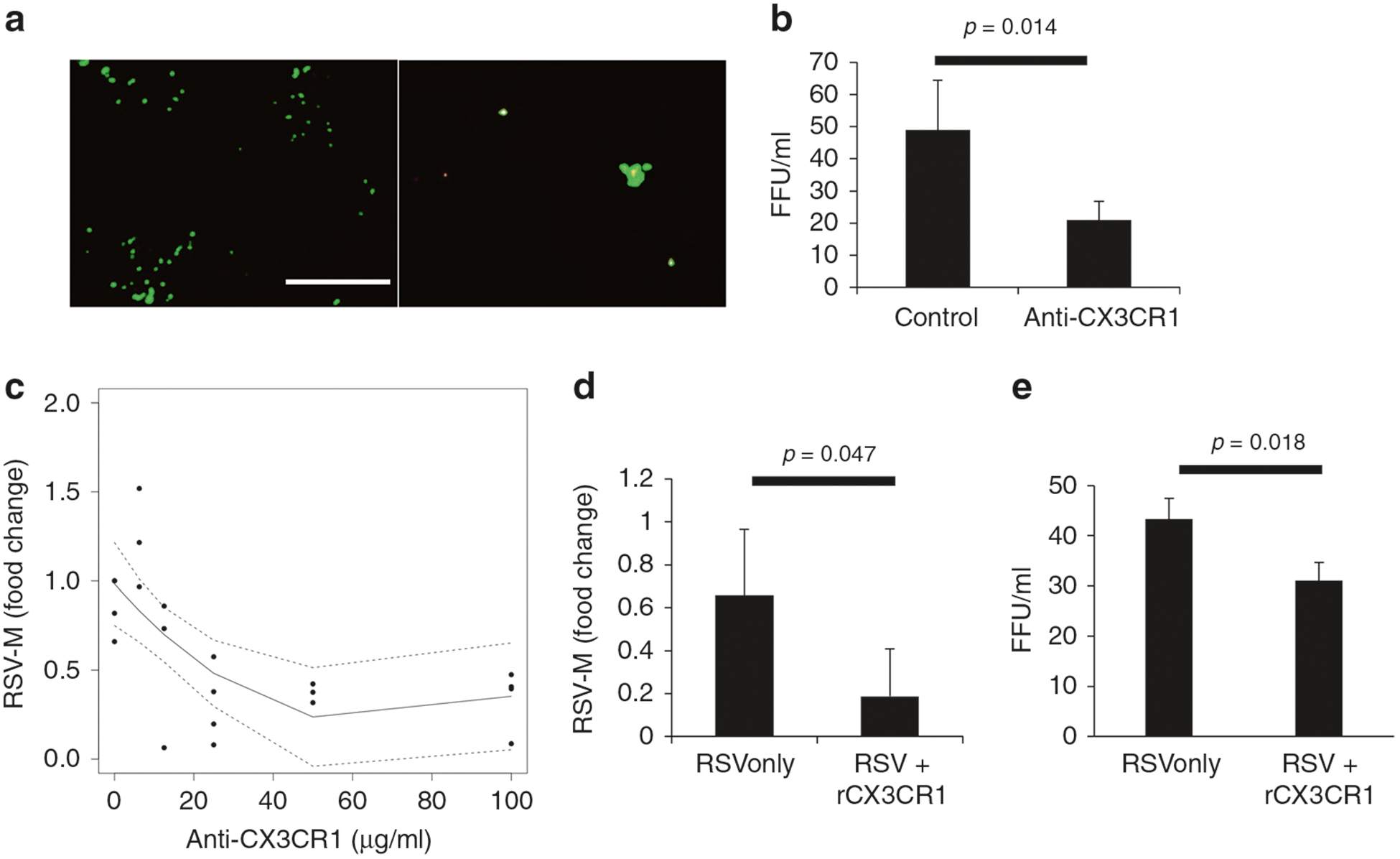
Fig. 4.

Effect of blocking CX3CR1 on RSV infection. a Representative fluorescent images of 24 h postinfection PHLE cell cultures infected with GFP-expressing RSV virus after cells were incubated for 30 min with (left) control antibody or (right) anti-CX3CR1 antibody. b Average RSV fluorescent focus-forming units per ml detected 24 h postinfection with or without CX3CR1-blocking antibody (25 μg/ml) (N = 4). c Dose–response curve of RSV quantification by qPCR of PHLE cell cultures infected with RSV after preincubation with varying levels of anti-CX3CR1 antibody (N = 4). d RSV M-protein transcript levels or e fluorescent foci 24 h postinfection with or without preincubation of RSV with recombinant CX3CR1 protein (N = 4).
