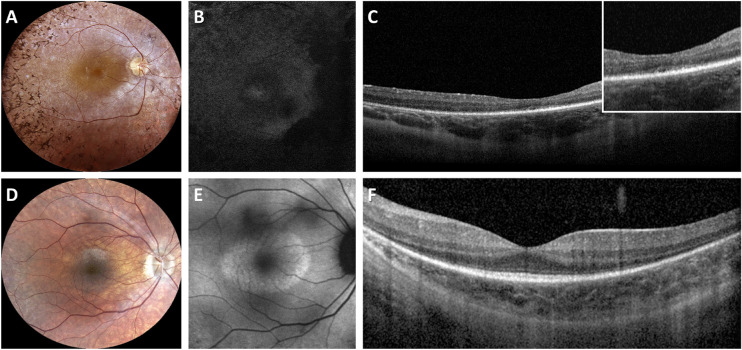
Figure 1.
Multimodal imaging findings in two representative patients with RPGR-associated RP. (A) Fundus photograph of a 31-year-old patient with typical RP (ID no. 13; BCVA 0,3; NM_001034853: c.1245+3A>T) showing bone-spicule pigment deposits in the mid peripheral retina, optic disc pallor and RPE atrophy. (B) Fundus autofluorescence (FAF) image showing a widespread hypoautofluorescence. (C) Spectral domain OCT revealing RPE atrophy. The inset shows the disruption of the ellipsoid zone (EZ) band. (D) Fundus photograph of a 25-year-old patient with sine pigmento RP (ID no. 37; BCVA 0,7; NM_001034853: c.1059+1G>A) showing absence of bone-spicule-like pigment migration, optic disc pallor, and mild RPE atrophy. (E) FAF image showing a hyperautofluorescent ring around the central macula. (F) Spectral domain OCT showing central sparing of the EZ band that is attenuated toward the peripheral macula.

