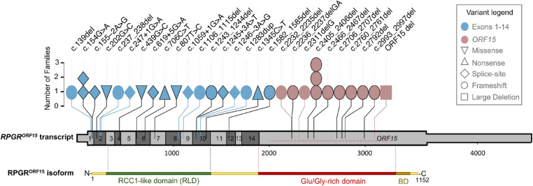
Figure 3.
Genetic variants detected in RPGR. Schematic drawing of the RPGRORF15 transcript showing the position and frequency of the variants identified in this cohort. The untranslated regions of the transcript are depicted as a thinner bar. Each symbol designates a family whose variant position is reported on the top. The symbol color indicates whether the variant is found in exons 1 to 14 or in the ORF15 region. The symbol shape indicates the mutation type. The large deletion (ORF15 del) is shown with a horizontal dotted line within the terminal exon of RPGRORF15. Already reported variants are denoted with a black border. Protein domains of the retina-specific isoform RPGRORF15 are shown below the transcript to indicate the RCC1-like domain (green), the Glu/Gly-rich region (red), and the basic domain (BD; brown; not to scale).

