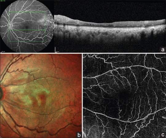
Figure 2.

(a) – SD-OCT image of the left eye through the area of retinal thinning predominantly involving the inner layers; (b) – multicolor image showing the retinal undulations corresponding to the area of retinal thinning and (c) – Composite OCTA image of the superficial and deep capillary plexus showing capillary dropout in the corresponding area
