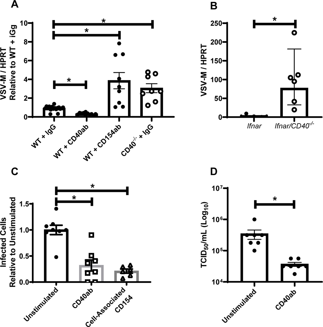
Figure 2. A BSL-2 model virus of EBOV, rVSV/EBOV GP, recapitulates EBOV findings.
A) WT or CD40−/− female mice were injected i.p. with 200 μg of agonistic CD40 antibody, blocking CD154 or IgG control as noted. Twenty-four hours later, mice were infected i.p. with a lethal dose of rVSV/EBOV GP. Peritoneal cells were isolated, RNA was harvested, and viral RNA was quantified by qRT-PCR at 24 hours of infection. B) Female Ifnar−/− and Ifnar/CD40−/− mice were infected i.p. with a dose of rVSV/EBOV GP that was lethal to Ifnar−/− mice. Twenty-four hours after infection, peritoneal cells were harvested, RNA was isolated and qRT-PCR analysis of viral RNA was performed. C-D) Enriched pmacs from male Ifnar−/− mice were incubated for 24 hours with agonistic CD40-specific mAb (C and D) or insect cell membranes expressing CD154 (C only). Treatments were removed and cells infected with rVSV/EBOV GP (MOI=1). Infection was quantified 24 hours following infection by flow cytometry and data are expressed as a percent change in the number of GFP+ cells relative to the unstimulated controls (C). Supernatants were titered on Vero cells (D). All experiments were performed three times. For all experiments, * indicates p<0.05.

