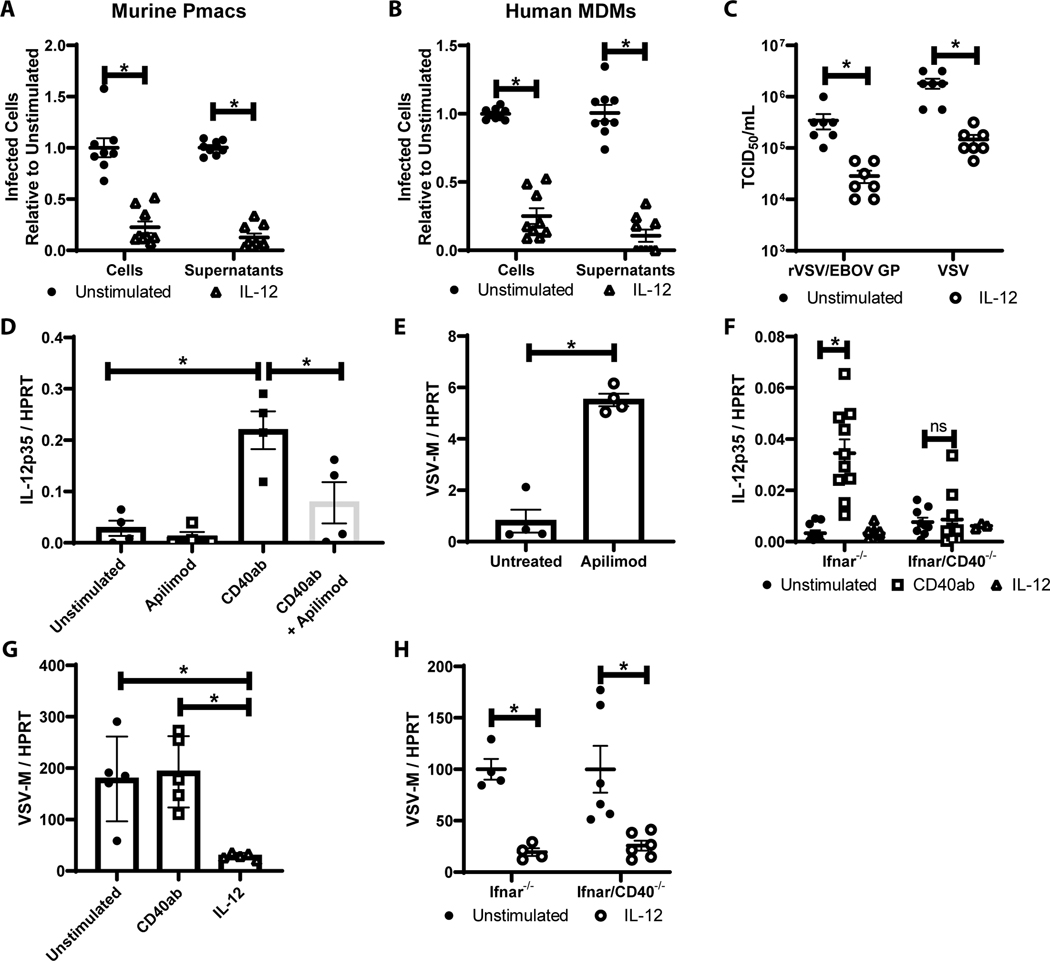
Figure 5. IL-12 production is a critical for CD40 restriction.
A/B) Peritoneal cells from male mice (A) or human monocyte derived macrophages (B) were untreated or treated with IL-12. After incubation, non-adherent cells were removed and enriched pmacs were infected for 24 hours with WT EBOV (MOI = 0.0015 pmacs; MOI = 0.1 MDMs) under BSL-4 containment. Cells were subsequently fixed, stained with anti-VP40 antibody and Hoescht dye, and infected cells were quantified by microscopy. Supernatants from infected cells were titered on Vero cells and quantified by the same microscopic method. C) Enriched pmacs from male Ifnar−/− mice were stimulated with IL-12 and at 24 hours infected with rVSV/EBOV GP (MOI=1) or VSV (MOI=0.5). Twenty-four hour following infection, supernatants were collected, filtered and TCID50s were determined on Vero cells. D/E) Pmacs were harvested from male Ifnar−/− mice and treated with agonistic CD40-specific mAb with or without the IL-12 inhibitor, apilimod, for 8 hours. RNA was harvested and qRT-PCR was performed to quantify IL-12p35 expression (D). In parallel, cells were stimulated with apilimod for 8 hours and infected with rVSV/EBOV GP (MOI=1). Twenty-four hours following infection, RNA was isolated and viral RNA was assessed by qRT-PCR (E). F) Enriched pmacs were obtained from Ifnar−/− and Ifnar/CD40−/− mice. Cells were stimulated with CD40 agonistic mAb or IL-12 and IL-12p35 gene expression was quantified by qRT-PCR. G) Pmacs from IL-12p40−/− mice were harvested and stimulated with agonistic CD40-specific antibody or IL-12 for 24h prior to infection with rVSV/EBOV GP (MOI=5). Twenty-four hours post infection, RNA was extracted and viral RNA was quantified by qRT-PCR. H) Enriched pmacs were obtained from Ifnar−/− and Ifnar/CD40−/− mice. Cells were stimulated with IL-12 and 24 hours later infected with rVSV/EBOV GP. Infections were evaluated for virus load by qRT-PCR at 24 hours. All experiments were performed three times. For all experiments, * indicates p<0.05.

