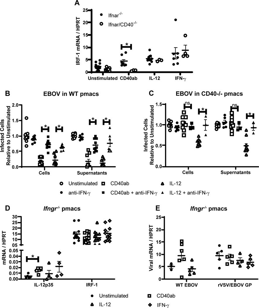
Figure 6. IL-12 stimulates IFN-γ that is required for CD40 restriction of virus replication.
A) Enriched pmacs were obtained from Ifnar−/− and Ifnar/CD40−/− mice. Cells were stimulated with CD40 agonist, IL-12 or IFN-γ and IRF-1 gene expression was quantified by qRT-PCR. B-C) Peritoneal cells from male WT (B) or CD40−/− (C) mice were incubated with agonistic CD40 mAb or IL-12 for 24 hours. Some cells were also treated with blocking anti-IFN-γ antibody as noted during antibody or cytokine treatment. After incubation, non-adherent cells were removed and enriched pmacs were infected with EBOV (Mayinga)(MOI = 0.0015) under BSL-4 conditions. At 24 hours of infection, cells were fixed, stained with anti-VP40 antibody and Hoescht dye, and infected cells were quantified by microscopy. In parallel, 24-hour supernatants from infected cells were titered on Vero cells and quantified by the same microscopic method. D-E) Enriched pmacs from male C57BL/6 Ifngr−/− mice were stimulated with the indicated stimuli. Twenty-four hours after stimulation, RNA was isolated from cells and IL-12p35 and IRF-1 mRNA was quantified by qRT-PCR (D) or infected with WT EBOV (MOI 0.0015) or rVSV/EBOV GP (MOI=5) and viral RNA was quantified 24 hours following infection by qRT-PCR (E). All experiments were performed three times. For all experiments, * indicates p<0.05.

