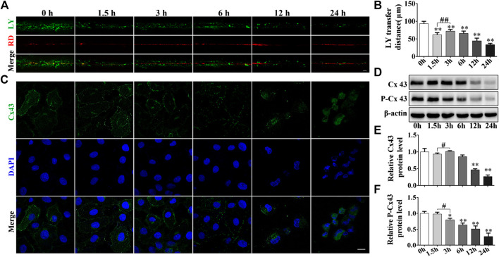
FIGURE 3.
Cd inhibits the GJIC in BRL 3A cells. (A) Following treatment with 5 μmol/L Cd for different time periods, GJIC was measured by the SL/DT method (scale bar = 50 μm). (B) Quantification of data obtained for the LY transfer distance depicting the spread of the dye from each side of the wound area (n = 6). (C) 5 μmol/L Cd disrupts Cx43 distribution in BRL 3A cells.Cx43 expression (green) and the presence of the nuclei (blue) were observed and imaged using confocal microscopy. Scale bar = 10 μm. (D–F) Cd treatment at different time periods affects the levels of connexin expression in BRL 3A cells. Compared with the control group, *p < 0.05, **p < 0.01; compared with the indicated group, # p < 0.05, ## p < 0.01.

