Figure.

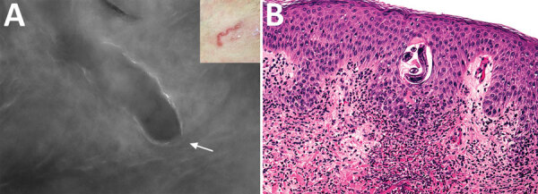
Imaging and biopsy results for patient with cutaneous larva migrans, Turin, Italy. A) Fluorescence-advanced videodermatoscopy showed larva with a diameter of 70–80 μm, located intraepidermally, ≈0.5 cm to the right of the distal end of the serpentine path (indicated in the inset) caused by its passage in the skin. White arrow indicates head of the larva (original magnification ×500). B) Hematoxylin and eosin–stained longitudinal skin section obtained from a 4-mm biopsy specimen, showing the cavity at the epidermal level in which it is possible to observe the larva inside (original magnification ×20). Once the biopsy specimen was obtained, confirmation of a larva by morphologic details or molecular techniques was necessary to differentiate animal nematodes from other larvae, particularly Strongyloides stercoralis, because treatment and follow-up would be different.
