Figure 1.
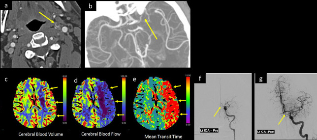
A 30-year-old male was tested positive for COVID-19 with marked deterioration of respiratory symptoms, suddenly developed aphasia and right-side-weakness with a low GCS. CTA of the head and neck, axial images at (a) suprahyoid neck and (b) at Sylvain fissure levels, show a floating thrombus within the cervical left ICA with intracranial extension occluding the left ICA terminus and M1 segment of left MCA (arrow in a and b). CTP of the brain post-processed (c) CBV image, (d) CBF, and (e) MTT color map images display large area of mismatched defect, with large perfusion defect along the left MCA territory showing decreased CBF, prolonged MTT, and compensated CBV (short arrows in c, d, and e respectively). Catheter angiography (f) before and (g) after successful thrombectomy with aspiration of the clot from left ICA show recanalization of left MCA and ACA. Unfortunately, few days later, the left ICA reoccluded. CBF, cerebral blood flow; CBV, cerebral bloodvolume; CTA, CT angiogram; CTP, CT perfusion; GCS, Glasgow coma scale; ICA, internalcarotid artery; MTT, mean transit time.

