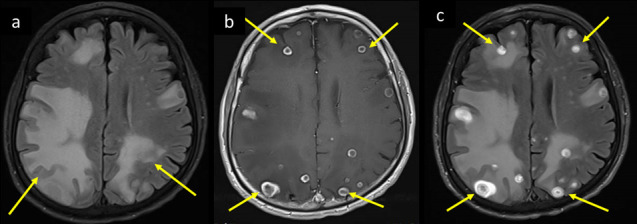
Figure 13.

A 65-year-old male, known heavy smoker with COPD presented to the ED with fever, increasing cough, severe headache, and brief episodes of left arm stiffness, face asymmetry, and aphasia. He rapidly progressed to status epilepticus with drop of GCS. He was intubated for airway protection and shifted to MICU, and started on antiepileptic medication. His PCR COVID-19 tested positive and CSF PCR was positive for TB. Brain MRI axial images at supraventricular level (a) T2-FLAIR, (b) corresponding T1WI post-i.v. contrast and (c) repeat T2-FLAIR post-i.v. contrast shows bilateral cerebral widespread vasogenic edema (long arrows in a–c), which surround ring enhancing lesions with central low signal intensity core (short arrows in a–c) representing extensive - flare up of tuberculomas. CSF, cerebrospinal fluid;COPD, chronic obstructive pulmonary disease; ED, emergency department; FLAIR, fluidattenuated inversion recovery; GCS, Glasgow coma scale; MICU, medical intensivecare unit; PCR, polymerase chain reaction; TB, tuberculosis.
