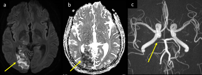
Figure 2.
A 58-year-old male diabetic with fever and mild respiratory symptoms was tested positive for COVID-19. He developed confusion, and headache increasing in severity over the past 2 days, associated with dizziness, blurring of vision, and left homonymous hemianopia. MRI (a) axial DWI b 1000, and (b) ADC map shows bright signal and corresponding low signal respectively along the right occipital lobe (arrow in a and b). Intracranial 3D MIP MRA (c) shows thrombotic occlusion of the P2 segment of right posterior cerebral artery (arrow in c). ADC, apparent diffusion coefficient; DWI, diffusion-weighted image; MIP, maximum intensity projection; MRA,MR angiography.

