Figure 5.

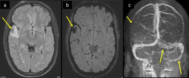
A 30-year-old male patient presented with recent episodes of tonic clonic seizures associated with post-ictal confusion. He had no fever, respiratory symptoms, or neck stiffness. He tested COVID positive by PCR. MRI brain, (a) axial FLAIR T2WI and (b) axial SWI shows right temporal intra-axial cortical and subcortical area of low signal on FLAIR T2WI with surrounding parenchymal bright signal edema, and blooming hypointensity in SWI images (arrow in a and b respectively). There was no diffusion restriction (not shown). (c) Source image of post-contrast MR venography shows filling defect in the torcula and left transverse sinus extending to the sigmoid sinus (long arrows in c), with abrupt cut-off of right superior anastomotic vein (short arrow in c). FLAIR, fluid attenuatedinversion recovery; PCR, polymerase chain reaction; SWI, susceptibility-weightedimaging.
