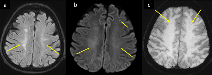
Figure 8.

A 59-year-old female with multiple comorbidities including DM, HTN, CKD, and paroxysmal AF, presented to the ED with 2 days history of shortness of breath, fever, and myalgia. She was tested positive for COVID-19, progressed to ARDS with altered level of consciousness and developed inability to move the left side of the body. MRI brain axial image at the supraventricular level (a) T2-FLAIR shows bilateral centrum semiovale white matter hyperintensities suggesting leukoencephalopathy (arrows in a). (b) Axial DWI b1000 shows corresponding facilitated diffusion of the white matter changes and subcortical lacunar infarcts of varying ages (long and short arrows respectively in b). (c) Axial SWI shows associated scattered punctate white matter blooming microbleeds (arrows in c). ARDS, acute respiratorydistress syndrome; AF, atrial fibrillation; CKD, chronic kidney disease; DM, diabetesmellitus; DWI, diffusion-weighted image; ED, emergency department; FLAIR, fluidattenuated inversion recovery; HTN, hypertension; SWI, susceptibility-weightedimaging.
