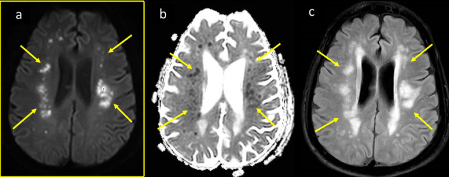
Figure 9.

A 56-year-old male COVID-19 positive patient with severe ARDS was intubated and admitted to ICU and was not waking up subsequently. MRI brain axial images at high ventricular level (a) DWI b1000, (b) corresponding ADC map, and (c) axial T2-FLAIR, show parasagittal white matter scattered chain of nodular (rosary beaded) diffusion restriction with high signal on DWI, low signal on ADC map (arrows in a, b), with more widespread T2-FLAIR white matter bright signal vasogenic edema (arrows in c). ADC, apparent diffusion coefficient; ARDS, acute respiratory distress syndrome; DWI, diffusion-weightedimage; FLAIR, fluid attenuated inversion recovery; ICU, intensive care unit.
