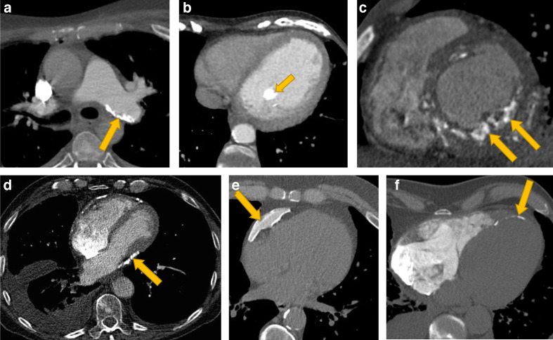
Figure 7.
Examples of cardiac calcification in other sites including (A) pulmonary arteries, (B) papillary muscle, (C, F) myocardium and (D, E) pericardium. Image (C) shows myocardial calcification in renal failure and (F) shows myocardial calcification in an infarct. (E) shows an example of benign pericardial calcification, whereas (D) shows pericardial calcification associated with constrictive pericarditis with associated atrial enlargement, tubular ventricular morphology, pleural effusion and a distended inferior vena cava.

