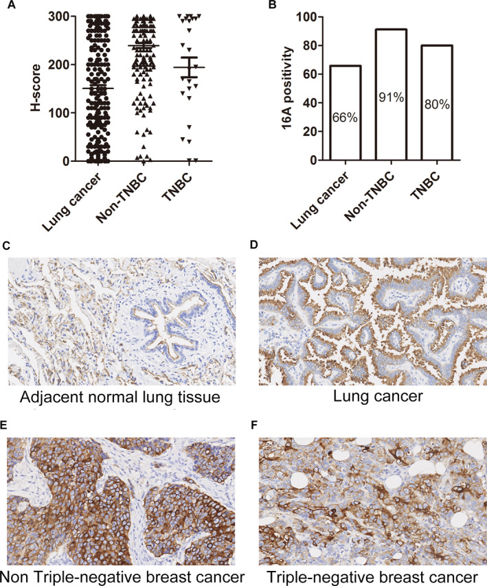
Figure 3.

Expression of the aberrantly glycosylated MUC1 peptide motif in lung and breast cancer tissues upon 16A antibody staining. (A) H‐score of the cancer tissue array stained with the 16A antibody. The intensity of IHC staining was scored at four levels: 0 (negative), 1 (weak staining), 2 (medium staining), and 3 (strong staining). The percentages of tumor cells at different intensity levels were evaluated. Total Score = (% at 0) ×0 + (% at 1) × 1 + (% at 2) ×2 + (% at 3) × 3. (B) 16A positivity in lung cancer, breast cancer, and triple‐negative breast cancer (TNBC) samples. (C, D, E, F) Representative photographs of MUC1 immunostaining in peritumoral (C), lung cancer (D), Non‐TNBC (E), and TNBC (F) tissue with the 16A antibody (original magnification ×200). Positivity was defined as ≥30% of tumor with staining ≥2+.
