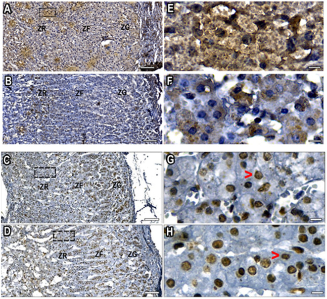
Figure 3.

DHEA/DHEAS immunostaining-positive cells at age 16 years (A and E), and at the age with a significant reduction in DHEAS blood level, at 43 years (B and F). Scale bar 100 µm (A and B) and 10 µm (E and F). ZR, zona reticularis; ZF, zona fasciculata; ZG, zona glomerulosa. Nuclei are shown in blue (hematoxylin counterstained), DHEA/DHEAS are stained brown. PTEN immunostaining-positive nuclei (brown) at peak DHEAS blood level at 19 years (C and G), and at the age with the most intense DHEAS blood level reduction, at 43 years (D and H). Scale bar 100 µm (C and D) and10 µm (G and H). The cytoplasmic PTEN signal was either not detectable in most cells or was a very weak signal in few cells (red arrow-heads). ZR, zona reticularis; ZF, zona fasciculata; ZG, zona glomerulosa.

 This work is licensed under a