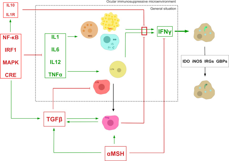
Fig 2. The ocular immunosuppressive microenvironment widely impairs the normal immune response to T. gondii infection.
In the general situation, the lysis of the parasitophorous vacuole and, ultimately, the parasite, relies on the expression of IFNγ by multiple cell types stimulated with various Th1 cytokines. In the eye, this mechanism is impaired by the presence of inhibitory molecules locally expressed by retinal cells, including RPE cells. Green arrows with pointy heads mean “activates/stimulates.” Red arrows with flat heads mean “inhibits.” αMSH, α-melanocyte-stimulating hormone; CRE, cAMP response element; GBP, interferon-induced guanylate-binding protein; IDO, indoleamine 2,3-dioxygenase; IL, interleukin; IFNγ, interferon γ; iNOS, nitric oxide synthases; IRF1, interferon regulatory factor 1; IRG, immunity-related guanosine triphosphatases; LT4, CD4+ T cells; LT8, CD8+ T cells; MΦ, macrophage; MAPK, mitogen-activated protein kinase; NF-κB, nuclear factor-kappa B; NKC, natural killer cells; PNL, polymorphonuclear leukocyte; TGFβ: transforming growth factor β; TNFα, tumor necrosis factor α; Treg, regulatory T cells.

